Effective Techniques for Coloring Animal Cell Diagrams: Labeled Animal Cell Coloring

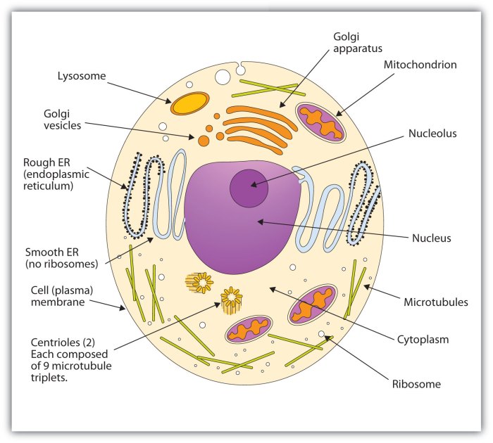
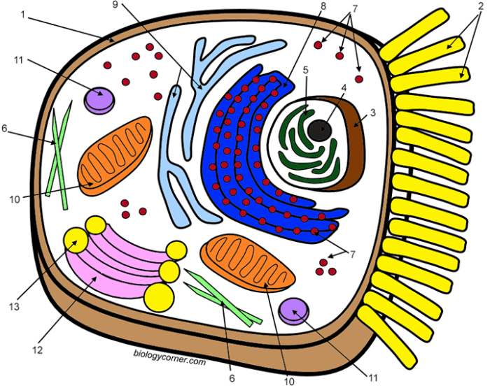
Labeled animal cell coloring – Coloring animal cell diagrams is a valuable educational tool for understanding cellular structure and function. By assigning different colors to organelles, students can visualize the distinct components of the cell and their relationships. This active learning approach enhances memory retention and promotes a deeper understanding of complex biological processes.Color-coding provides a visual method for differentiating between the various organelles and their functions within the animal cell.
This technique allows students to quickly identify and associate specific colors with particular organelles, making it easier to grasp the overall organization of the cell.
Color-Coding Organelles and Their Functions, Labeled animal cell coloring
The strategic use of color in diagrams helps highlight the roles of various organelles. For instance, using a vibrant color like red for the mitochondria emphasizes its role as the powerhouse of the cell, generating energy. Similarly, using green for the Golgi apparatus can link it visually to its function in processing and packaging proteins, much like a distribution center.
Step-by-Step Guide for Effective Coloring
Following a structured approach ensures clear and informative diagrams. This step-by-step guide Artikels an effective coloring process:
- Start by choosing a light color for the cytoplasm, such as a light blue or pink. This provides a neutral background that makes the other organelles stand out.
- Select contrasting colors for the nucleus and nucleolus. For example, use a darker blue for the nucleus, which houses the genetic material, and a contrasting color like purple for the nucleolus, the site of ribosome production.
- Color the endoplasmic reticulum (ER) with a color that suggests its interconnected network. A light purple or lavender can represent the smooth ER, while a slightly darker shade can distinguish the rough ER, dotted with ribosomes.
- Use a vibrant color, such as red or orange, for the mitochondria to highlight its role in energy production.
- Choose a color like green for the Golgi apparatus to visually represent its processing and packaging functions.
- Color the lysosomes, responsible for cellular digestion, with a darker color like brown or deep red.
- Use a distinct color for the cell membrane, such as yellow or orange, to emphasize its role as the outer boundary of the cell.
- Finally, add a key or label to your diagram that clearly identifies each organelle and its corresponding color. This allows for easy reference and reinforces the association between color and function.
Example Color Palette
A sample color palette could include: light blue for cytoplasm, dark blue for the nucleus, purple for the nucleolus, light purple for smooth ER, darker purple for rough ER, red for mitochondria, green for Golgi apparatus, brown for lysosomes, and yellow for the cell membrane.
Benefits of Using a Color Key
A color key is crucial for interpreting the colored diagram. It provides a clear legend that connects each color to a specific organelle, making the diagram easy to understand. For instance, a key might show a red square labeled “Mitochondria,” a green square labeled “Golgi Apparatus,” and so on. This eliminates ambiguity and ensures that the information conveyed is accurate and accessible.
Variations and Adaptations of Animal Cell Coloring

Animal cell coloring offers a dynamic approach to understanding cell structure and function. Its adaptability across various learning levels and styles makes it a valuable tool in biological education. By modifying complexity and integrating interactive activities, educators can tailor the coloring experience to suit individual learning needs and objectives.Coloring diagrams provides a visual and kinesthetic learning experience that aids in memory retention and comprehension.
This section explores how adjustments to animal cell coloring activities can cater to different educational contexts.
Simplified and Complex Animal Cell Diagrams
Animal cell diagrams exist in a spectrum of complexity. Simplified versions typically present the major organelles, such as the nucleus, cell membrane, and cytoplasm. These basic diagrams are suitable for younger learners or introductory biology lessons, providing a foundation for understanding basic cell structure. More complex illustrations incorporate a wider range of organelles, including the Golgi apparatus, endoplasmic reticulum, ribosomes, mitochondria, lysosomes, and centrioles.
These detailed diagrams are appropriate for advanced students exploring cellular functions in greater depth.
Integrating Learning Activities with Animal Cell Coloring
Animal cell coloring can be enhanced by combining it with other learning activities. Labeling exercises reinforce the association between organelle names and their visual representations. Students can label pre-colored diagrams or label their own colored diagrams. Research projects can delve into the specific functions of each organelle. Students can research and present information on the roles of organelles in cellular processes, connecting the visual representation to its biological significance.
Creating 3D models using clay or other materials can further enhance spatial understanding of cell structure.
Adapting Animal Cell Coloring for Different Learning Styles and Age Groups
Animal cell coloring can be adapted for various learning styles and age groups. For younger learners, coloring can be combined with storytelling or simple matching activities. For visual learners, color-coding organelles based on their functions can be beneficial. Kinesthetic learners might benefit from creating their own diagrams or building 3D models. For older students, coloring can be integrated with more complex activities such as designing presentations or creating interactive digital models.
For example, students could research a specific disease related to a cellular malfunction and present their findings alongside a colored diagram highlighting the affected organelle. Another adaptation could involve students creating a color-coded chart linking each organelle to its function, along with a mnemonic device to aid memorization.
Exploring the intricate details of labeled animal cell coloring can be fascinating. For a fun change of pace, try some safari animals coloring pages to explore the exterior beauty of these complex organisms. Then, return to your animal cell diagrams with a renewed appreciation for the inner workings that give these magnificent creatures life.
